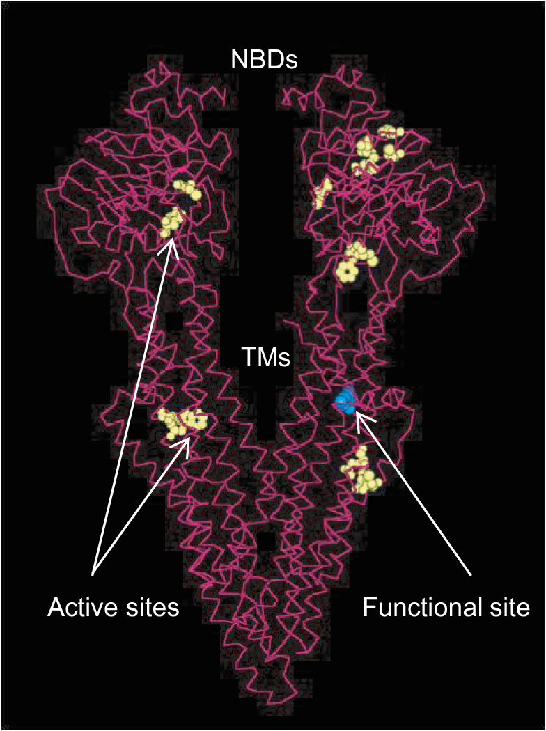
Fig. 8.
Protein structure simulation of qph1 generated using Pymol. The six active sites of the protein are shown in yellow; the Arginine to Leucine amino acid substitution on the ninth α-helix in the transmembrane domain is indicated in blue. TMs, transmembrane domains; NBDs, nucleotide binding domains.

