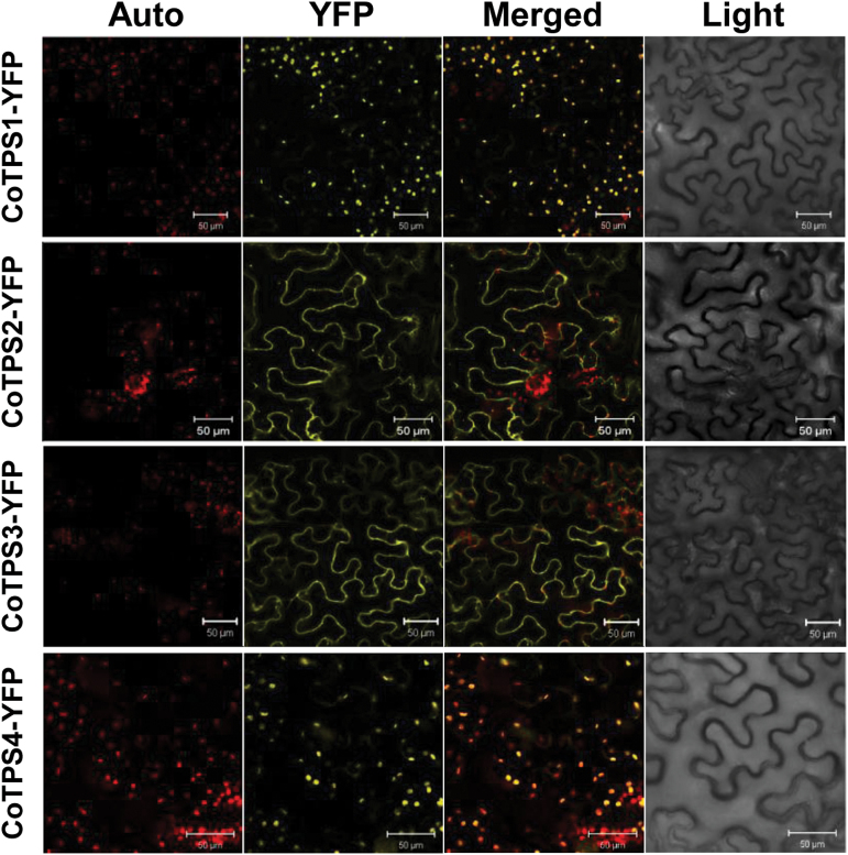
Fig. 5.
Subcellular localization of CoTPSs. YFP-fused CoTPSs (CoTPS1–YFP, CoTPS2–YFP, CoTPS3–YFP, and CoTPS4–YFP) were transiently expressed in N. benthamiana leaves by Agrobacterium-mediated infiltration and visualized at 3 d post-infiltration using the YFP channel of a confocal microscope. Auto, chlorophyll autofluorescence; YFP, YFP channel image; Light, light microscopy image; Merged, merged image between Auto and YFP. Bars, 50 µm. (This figure is available in colour at JXB online.)

