
Figure 1.
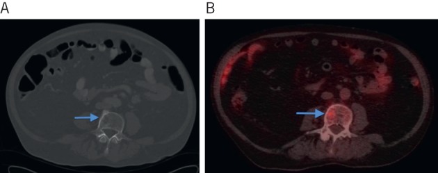
Computed tomography (CT) showing subtle abnormality in the L3 vertebral body (A) and positron emission tomography CT showing increased avidity in the same region in keeping with a metastasis (B).
Official websites use .gov
A
.gov website belongs to an official
government organization in the United States.
Secure .gov websites use HTTPS
A lock (
) or https:// means you've safely
connected to the .gov website. Share sensitive
information only on official, secure websites.
Computed tomography (CT) showing subtle abnormality in the L3 vertebral body (A) and positron emission tomography CT showing increased avidity in the same region in keeping with a metastasis (B).