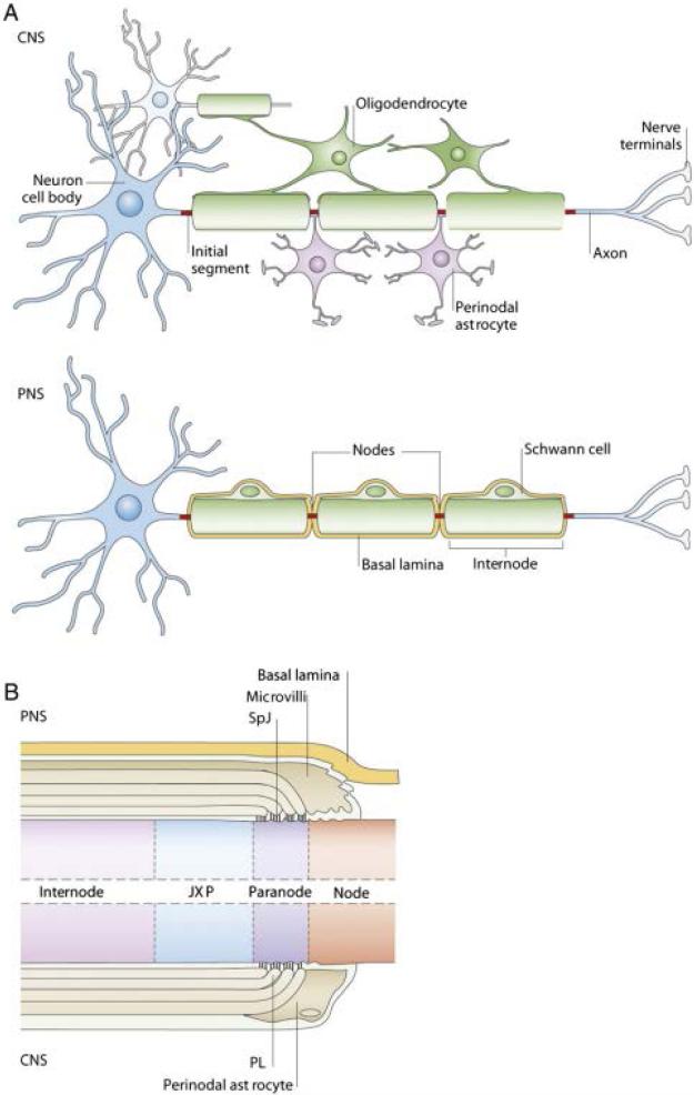
Figure 1.
Illustration modified from Poliak and Peles et al. [11] showing the differences between myelinated fibers in the Peripheral Nervous System (PNS) and the Central Nervous System (CNS). A. Oligodendrocytes myelinate the CNS wrapping their processes around multiple axonal segments. Schwann cells are the myelinating cell type in the PNS and contact single axonal segments. Discontinuities of the myelin sheath along the axon known as nodes of Ranvier are contacted by perinodal astrocytes in the case of the CNS, whereas in the PNS Schwann cells extend microvilli to the node that is surrounded by basal lamina, highlighting the multifunctional capacity of these cells. B. Representation of a longitudinal cut of the myelinated fiber surrounding a node of Ranvier in the PNS (top) and CNS (bottom). The paranode, adjacent to the nodes, is formed by non-compact myelin and contains reflexive gap junctions establishing a communication compartment across the membranous myelin layers. The juxtaparanode (JXP) is composed of compact myelin and divides the paranodal region of the internodal region.

