Fig. 6.
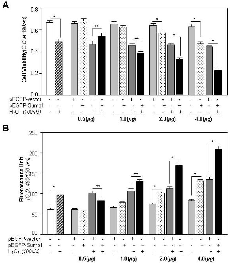
Sumo1 overexpression reduced cell viability and increased the ROS level in concentration-dependent fashion at normal physiological condition, and LECs were more susceptible to oxidative stress. hLECs were overexpressed with different concentrations of pEGFP-vector or pEGFP-Sumo1 and pcDNA3 plasmid. A required amount of pcDNA3 plasmid was used to equalize DNA load during transfection. Complete media were replaced with 0.2% BSA, and cells were exposed to H2O2 (100μM). After 24h, cell viability (A) and ROS level (B) were examined. Histogram values represent mean ± SD of three independent experiments (**p<0.05; *p<0.001 vs control).
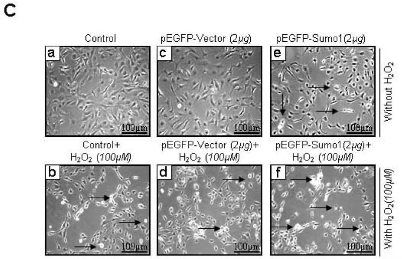
(C) Cells overexpressing Sumo1 were more susceptible to cell death in response to oxidative stress. Photomicrographs of cells showed relative cell death (white rounded cells, indicated by arrow and empty spaces where cells detached after death) in cells overexpressing Sumo1. hLECs (8 × 10⁵) were transfected with pEGFP-vector or pEGFP-Sumo1 and exposed to H2O2 (100μM). 24h later cells were photomicrographed. a, control (no transfection); b, control+H2O2 (100μM); c, pEGFP-Vector (2μg); d, pEGFP-Vector (2μg)+H2O2 (100μM); e, pEGFP-Sumo1(2μg); f, pEGFP-Sumo1(2μg)+ H2O2 (100μM).


