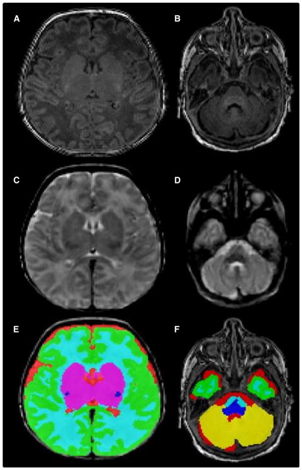
Figure 1.
Input and output for the automatic segmentation of brain tissue volumes. A, Axial T1 slice at the level of the thalami and B, cerebellum; C, axial T2 slice at the level of the thalami and D, cerebellum; E, automatic segmentation of brain tissue volumes at the level of the thalami and F, cerebellum. The tissue classes depicted are CGM (green), unmyelinated white matter (light blue), mWM (dark blue), SCGM (magenta), CSF (red), and cerebellum (yellow).

