
FIG 1.
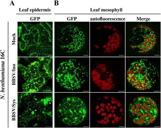
BBSV infection induced severe morphological changes in the endoplasmic reticulum. BBSV virions were inoculated onto N. benthamiana line 16C, in which the ER is decorated with GFP (37). The treatment is indicated on the left of each row. Both leaf epidermis (A) and mesophyll (B) cells were analyzed by CLSM. N. benthamiana 16C leaves were inoculated with BBSV virions (BBSV/Ino). At 3 dpi, the leaf epidermis was peeled from these leaves, and the remainder was treated with enzymes to liberate protoplasts from the mesophyll. Systemically infected leaves (BBSV/Sys) were harvested at about 8 dpi and similarly processed for CLSM analysis. Mock-inoculated leaves (Mock) from line 16C were processed following the same procedures. The arrowheads indicate the ER aggregates. Bars = 10 μm.