FIG 2.
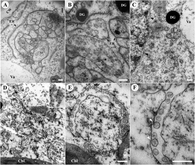
BBSV remodels the ER membranes. TEM was carried out to analyze the cytopathological changes during BBSV infection of N. benthamiana. (A) BBSV infection led to ER aggregation and large amounts of viral particles intermingled within the convoluted ER membrane. The arrow indicates the ER. (B to F) BBSV-induced vesiculation of the ER and VPs were often present in the dilated ER cisternae. (B) VPs were observed in the aggregates of branched ER cisternae (⭑), and a virus crystal (Vi) was in proximity to the VPs. Electron-dense globules (DG) were occasionally visible in the cytoplasm. The arrow indicates the ER. (C) Different numbers of spherules were enclosed in the VPs, and sometimes spherules appeared in the lumen of the perinuclear membrane (arrowheads). (D) Some spherules seem to be suspended in the cytoplasm (arrowheads). The enlarged image (top right corner) shows the region pointed to by the arrowhead. (E) Higher magnification of an area in panel D (dashed rectangle) shows the presence of fine fibrils in the VPs. (F) Membrane-associated spherules (further enlargement of the dashed rectangle in panel E) showing potential connections between the spherules and the outer ER membrane (arrowheads). Chl, chloroplast; Vi, virus particles or virus crystals; Mit, mitochondria; CW, cell wall; Nu, nucleus; Va, vacuole. Bars, 200 nm.

