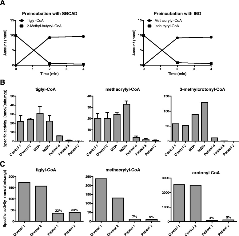
Fig. 5.

SCEH enzyme activity in patient fibroblasts measured with different substrates. a Formation of tiglyl-CoA by incubation of 2-methyl-butyryl-CoA with recombinantly expressed short branched-chain acyl-CoA dehydrogenase (SBCAD) (left panel) and formation of methacrylyl-CoA by incubation of isobutyryl-CoA with isobutyryl-CoA dehydrogenase (IBD) (right panel). After 2 min all substrate is converted into product. b Enoyl-CoA hydratase enzyme activity with tiglyl-CoA (left graph), methacrylyl-CoA (middle graph) and 3-methylcrotonyl-CoA (right graph) in fibroblasts of two control subjects, SCEH deficient patients and a patient with a complete deficiency of mitochondrial trifunctional protein (MTP) and a patient with 3-methylglutaconyl-CoA hydratase (MGH) deficiency. c Enoyl-CoA hydratase enzyme activity with tiglyl-CoA (left graph), methacrylyl-CoA (middle graph) and crotonyl-CoA (right graph) in liver of two control subjects and two SCEH deficient patients (patient 1 and 2). Residual activity in patients samples is indicated in % of mean activity in two control liver samples
