Figure 2.

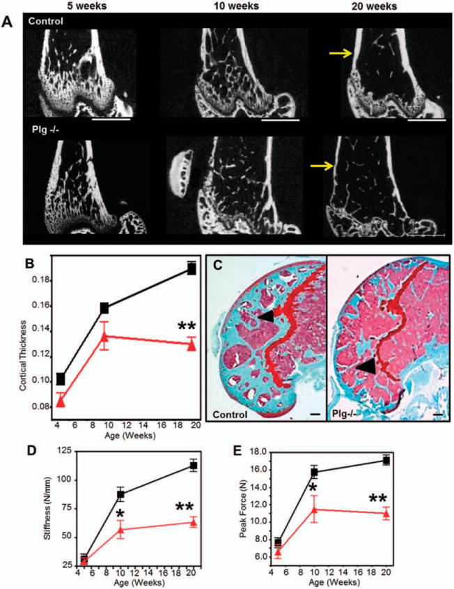
Plasminogen-deficient (PLG−/−) mice develop early degeneration and reduced fracture resistance of the appendicular skeleton. Triangles represent PLG−/− mice; squares represent control mice. A, Two-dimensional images rendered from micro–computed tomography (micro-CT) data reveal cortical thinning in PLG−/− mice compared to control mice (arrows) occurring at age 20 weeks despite relatively normal bone architecture in younger mice. Bars = 1 mm. B, Shown are tabulated cortical measurements (Student’s t-test was performed for each time point using data collected from separate mice at the time that they were killed). C, Safranin O staining of the distal femur at age 20 weeks demonstrates loss of trabecular bone in the distal femoral epiphysis in PLG−/− mice compared to control mice (arrowheads) (see Supplementary Table 1, available on the Arthritis & Rheumatology web site at http://onlinelibrary.wiley.com/doi/10.1002/art.38639/abstract). Bars = 200 μm. D and E, Biomechanical analyses of stiffness (D) and fracture resistance by peak force (E) indicate that observed progressive loss of bone results in significantly reduced biomechanical strength (means, standard deviations, and sample sizes, along with additional biomechanical and micro-CT data, are presented in Supplementary Tables 1 and 2, available on the Arthritis & Rheumatology web site at http://onlinelibrary.wiley.com/doi/10.1002/art.38639/abstract). Bars show the mean ±SD (n = 8 wild-type mice; n=11 PLG−/− mice). *=P<0.05; **=P<0.01 versus control mice, by Student’s t-test. Color figure can be viewed in the online issue, which is available at http://onlinelibrary.wiley.com/doi/10.1002/art.38639/abstract.
