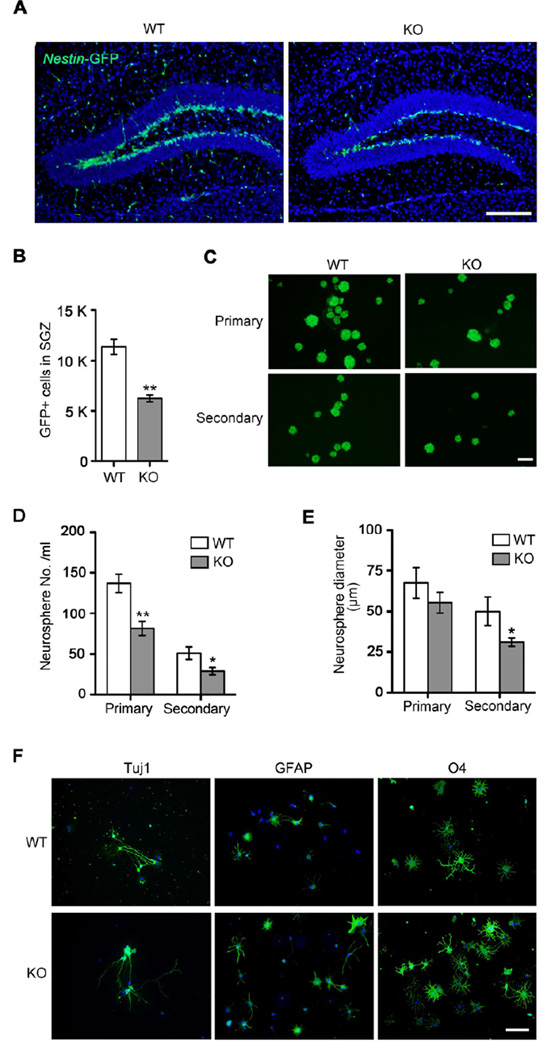
Figure 2. Reduction of Nestin-GFP Positive Neural Progenitor Cells in the Tet1 Deficient Dentate Gyrus and Their Impaired Proliferation in Vitro.
(A) Observation of neural progenitor cells in the adult SGZ using Nestin-GFP transgenic mice. Shown are coronal images of the SGZ in 4-month-old WT (Tet1+/+; Nestin-GFP) and KO (Tet1−/−; Nestin-GFP) mice captured at the same exposure. Scale bar, 100 µm.
(B) Quantification analysis of Nestin-GFP positive cells in the subgranular cell layer of dentate gyrus (n = 3 pairs of mice).
(C) Isolation and culture of Nestin-GFP positive progenitors from 2-month-old WT and KO dentate gyrus by FACS. Scale bar, 100 µm.
(D) Quantification of primary and secondary neurospheres (The initial seeding is 20,000 cells/ml, n = 3 cases).
(E) Average diameters of primary and secondary neurospheres (n = 3 cases).
(F) The tripotent differentiation capacity of Tet1-deficient NPCs. Neurons, astrocytes and oligodendrocytes were induced by in vitro differentiation of neurospheres isolated from WT and Tet1 KO DG. Cell lineage markers used: Tuj1 for neurons, GFAP for astrocytes and O4 for oligodendrocytes. Scale bar, 100 µm.
All quantifications are presented as mean ± s.e.m. and analyzed by two-tailed t-test. ** P < 0.01, * P < 0.05.
See also Figure S1.

