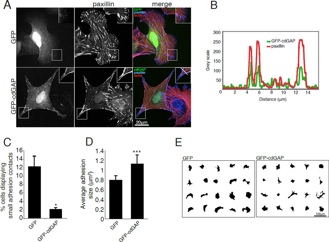
Figure 4. CdGAP over expression inhibits the formation of small adhesion contacts and increases adhesion area.
(A) U2OS cells transfected with GFP or GFP-cdGAP were spread on 10µg/ml collagen coated coverslips and stained for paxillin and actin. Inset shows the localization of GFP-cdGAP to adhesions that are positive for paxillin staining. The white bar in the merged inset for the GFP-cdGAP expressing cell marks adhesions through which the line profile in (B) was drawn. (B) A line profile through adhesions demonstrates that GFP-cdGAP and paxillin fluorescence intensity peaks in adhesions and diminishes in the surrounding cytoplasm. (C) The percentage of cells with small leading edge or peripheral adhesions decreased in GFP-cdGAP expressing cells. The percentage of small adhesions was quantified from a minimum of 60 transfected cells from each of three independent experiments per condition and GFP versus GFP-cdGAP expressing cells compared with Student’s t-test, *, p < 0.05. (D) Average adhesion size was measured in U2OS cells expressing GFP and GFP-cdGAP constructs from a total of 30 transfected cells per condition from three independent experiments and compared using Student’s t-test, ***, p < 0.001. (E) Masks of cells over expressing GFP or GFP-cdGAP demonstrate that GFP-cdGAP over expressing cells have a smaller spread area and an angular morphology. For Figure 4, all error bars represent +/− the standard error of the mean.

