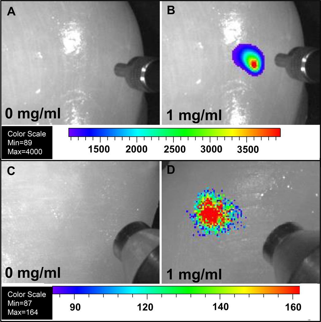
Figure 8.
Deep tissue imaging of radioluminescent and upconversion nanophosphors following injection 1 cm deep into chicken breast for 200 µl of (A) PBS solution under 980 nm laser, (B) 1 mg/mL nanophosphors with encapsulated MTX under 980 nm laser, (C) PBS solution under X-ray, (D) 1 mg/mL nanophosphors with encapsulated MTX under X-ray. The concentric cylindrical object in Figure A and B at the right of each image is a SMA905 terminus of an optical fiber used to illuminate the tissue with 980 nm light. The concentric cylindrical object in Figure C and D at the right of each image is a mini-X-ray tube.

