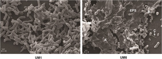
Fig. 4.

Scanning electron microscope micrograph of biofilm formation by B. pseudomallei on a glass slide. B. pseudomallei UM1demonstrated reduced biofilm formation compared to UM6. a Extracellular polymeric substance (EPS) is clearly visible around the B. pseudomallei UM6 colonies. b The matrix is holding the bacteria together but has not yet been encapsulated. c Pilus protruding from a UM6 colony
