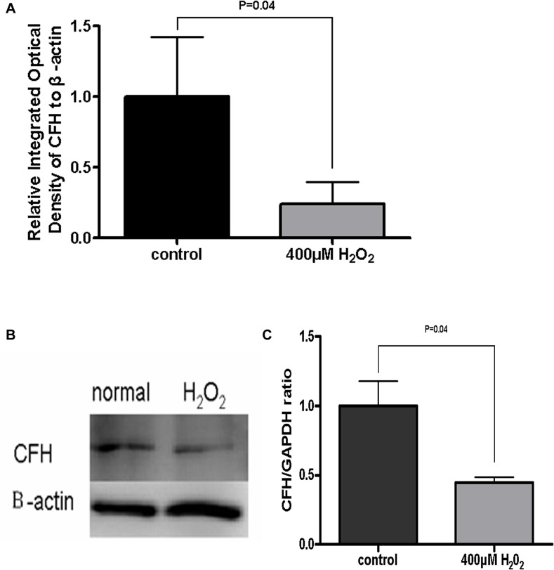
Fig 4. CFH expression reduced in the H2O2 treated ARPE-19 cells.
Real-time PCR showed that the CFH mRNA expression in H2O2-damaged ARPE-19 cells was significantly lower than that in control cells (P < 0.05) (A). In Western blots, the CFH band of H2O2-damaged ARPE-19 cells was paler than the normal cells (B). The bands’ gray scale was quantified by Quantity One software. Compared to control cells, the CFH expression was reduced significantly in H2O2-damaged ARPE-19 cells (P < 0.05) (C). Data are presented as mean ± SD from results done with three independent biological samples, each analyzed with triplicates.

