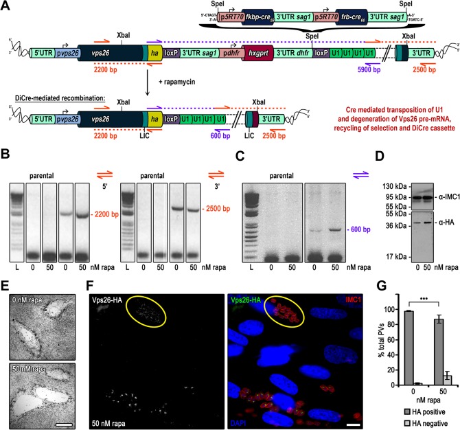
Fig 4. Targeted silencing of vps26 in Δku80-DHFR T. gondii parasites.
(A) Schematics of the genomic locus of vps26 after single homologous integration of the XbaI linearised endogenous ha tagging and U1 gene silencing construct in absence and presence of rapamycin. A codon optimised DiCre cassette is cloned into the SpeI restriction site between the hxgprt selection cassette and the second loxP site. (B) and (C) Analytical PCR on genomic DNA extracted from clonal VPS26-HA-U1 parasites and the parental line Δku80-DHFR grown for 24 h in absence and presence of 50 nM rapamycin. (B) Construct integration was confirmed by using oligos indicated as orange arrows in (A). Theoretical fragment sizes of 2200 bp and 2500 bp for 5’ and 3’ integration were amplified respectively in VPS26-HA-U1 parasites independent of rapamycin. (C) Confirmation of Cre/loxP site specific recombination. Binding sites of primers used for analysis are indicated with purple arrows in (A) with the theoretical fragment sizes. Even though it was impossible to amplify the very large spacer (5900 bp) in absence of rapamycin the PCR product of 600 bp in presence of rapamycin confirms Cre/loxP site specific recombination. L, ladder. (D) Immunoblot analysis of clonal VPS26-HA-U1 parasites cultured for 48 h with or without 50 nM rapamycin. Membrane probed with anti-HA and anti-IMC1 as loading control. In presence of rapamycin no downregulation of VPS26-HA expression was observed. (E) Giemsa stain. Growth analysis over a 7 days period in absence and presence of 50 nM rapamycin shows no difference in plaque formation. Scale bar represents 500 μm. (F) Immunofluorescence analysis of clonal VPS26-HA-U1 parasites grown for 48 h in presence of 50 nM rapamycin. Green, HA; red, IMC1; blue, DAPI; scale bar represents 10 μM. Endogenous HA-tagged VPS26 localises apical to the nucleus in Golgi region. Parasites with silenced vps26 are encircled in yellow. (G) Quantification of vps26 silencing efficiency. The graph shows the percentage of HA positive and negative vacuoles determined by examination of 100 vacuoles per condition based on immunofluorescence analyses of clonal VPS26-HA-U1 parasites grown for 48 h in absence or presence of 50 nM rapamycin. Values are means ±SD (n = 3). Under rapamycin conditions a vps26 downregulation of 12.6 ± 5.32 was obtained (***, p<0.001, Mann-Whitney test).

