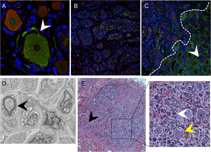
Fig 2. DRG architecture is maintained in DRG xenografts.
Panels A-C, DRG xenograft, 20 weeks after transplantation, stained with subtype specific markers anti-RT97 (green)/anti-peripherin (red) antibody (A-C). White arrow in (A) denotes the axon hillock at the neuronal cell body. White dotted line in (C) delineates the margin between the DRG xenograft and the murine kidney, arrow shows axons projecting into the murine kidney. Panel D, transmission electron micrograph of DRG xenograft with arrow showing myelinated nerve fiber. Panel E, DRG xenograft 80 weeks after implantation. Black arrow in (E) denotes the nerve root, panel on the right (F) is inset panel (black box) from (E) with arrows showing small, dark neurons (white) and large, light neurons (yellow).

