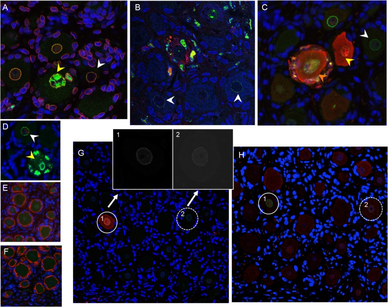
Fig 5. VZV restriction in mechanoreceptive neurons occurs after viral entry.
For each panel, one representative image is shown for staining experiments performed on multiple tissue sections (6–10 sections) for each DRG. (A-C) ORF23 capsid protein is a marker of virion entry as well as the later formation of progeny virions; virion entry is indicated by ORF23 capsid puncta at the nuclear rim (green), co-stained with cellular lamin A/C (A, red), and prior to expression of IE62 (B, red) or IE63 (C, red). (D) Dual staining for ORF23 (green) and cellular PML (red). (E) ORF23 nuclear rim staining was not observed when using pre-immune serum (green); red is N-CAM staining. (F) ORF23 capsid protein is not detected in uninfected DRG, as shown by staining for N-CAM (red) and absence of ORF23 (green). (G-H) Staining of adjacent tissue sections stained with antibody for ORF23 (green) and IE63 (G, red) or RT97 (H, red). Two neurons are circled and shown in both panels. The single 488-channel images for Panel G, shown in greyscale for better visualization, are provided for the circled neurons (1 and 2). The contrast of the G, neuron 2, ORF23 B&W panel, is enhanced so that the discrete ORF23 particles are easier to observe.

