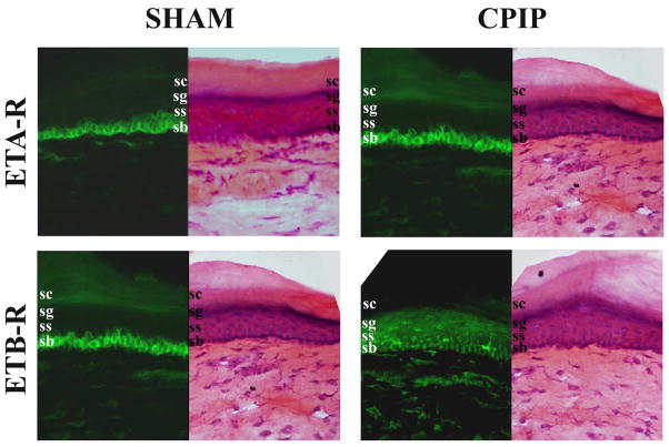
Fig. 5. Distribution of ETA-R and ETB-R in the skin of sham and CPIP mice.
Two days post I/R injury, the skin of sham (left panels) and CPIP (right panels) mice was incubated with anti-ETA-R (1:4000, top panels, green) or anti-ETB-R (1:2000, bottom panels, green) antibodies, or stained with hemathoxiline and eosin (H&E). All pictures were taken with a 40 X objective. ETA-R staining was predominantly found in the deeper stratum basalis (s.b.) layer and ETB-R in the medium strati granulosum (s.g.) and spinosum (s.s.) layers. No or poor staining was observed in the external stratum corneum (s.c.) There are no obvious changes in ETA-R or ETB-R staining between sham and CPIP mice.

