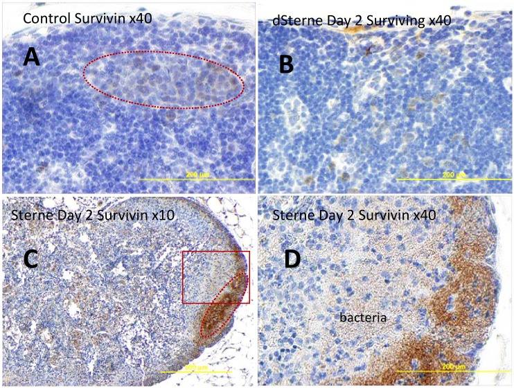
Fig 8. Immunohistochemical staining of formalin-fixed sections of popliteal LNs for survivin (brown) in naïve (A) and B. anthracis Sterne-challenged mice (B-D) at different magnifications.
Hematoxylin counterstain (blue) was used. The majority of survinin+ cells (immunostained brown) localize to the cortical zones morphologically similar to B-cell lymphoid follicles (dotted lines in A and C) overlapping with bacteria in infected mice (bottom panels C, D). The boxed region in the left bottom panel C is magnified on the right.

