
Fig 4. Both IL-10 and GC facilitate the phosphorylated-p38 MAPK nuclear translocation in Caco-2 cell monolayers.
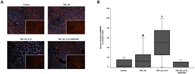
Panel A shows the nuclear arrangement of phosphorylated p38 MAPK in cultured Caco-2 cell monolayers, by immunofluorescence staining with anti-phospho-p38 MAPK (Thr180/Tyr182) (red) and Hoechst dye (blue). Scale bar 20 μm. White squares show images without Hoechst staining. Panel B shows the median (IQR, limits) percentage of cells with positive phospho-p38 MAPK nuclear staining. *p≤ 0.021 vs. other groups, #p≤ 0.043 vs. TNF_GC_IL-10_SB203580.