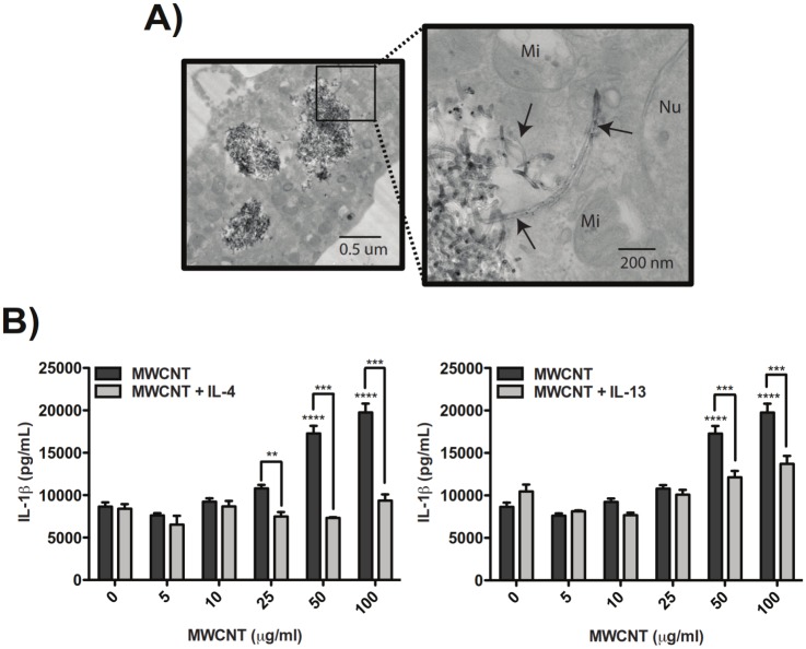
Fig 1. MWCNT-induced IL-1β secretion by LPS-primed THP-1 cells is inhibited by Th2 cytokines in vitro and corresponds to alternative macrophage activation.
(A) Transmission electron micrographs demonstrating phagocytosis of MWCNTs by THP-1 cells after exposure to 10 μg/mL MWCNT for 24 hours (Mi = mitochondria, Nu = nucleus). Images taken at 14000X and 56000X, respectively. (B) Secreted IL-1β measured by ELISA in supernatants from LPS-primed THP-1 cells after exposure to MWCNTs for 24 hours and suppression of MWCNT-induced IL-1β secretion by co-incubation with 10 ng/ml IL-13 or IL-4. Data for MWCNTs alone is the same for both graphs. Statistical analysis was performed using a one-way ANOVA with a post hoc Tukey. ****P < 0.0001 for MWCNT treatment compared to untreated control. ***P < 0.001, **P < 0.01 for IL-13 or IL-4 co-exposure compared to MWCNTs alone. Graphs are representative of multiple experiments.

