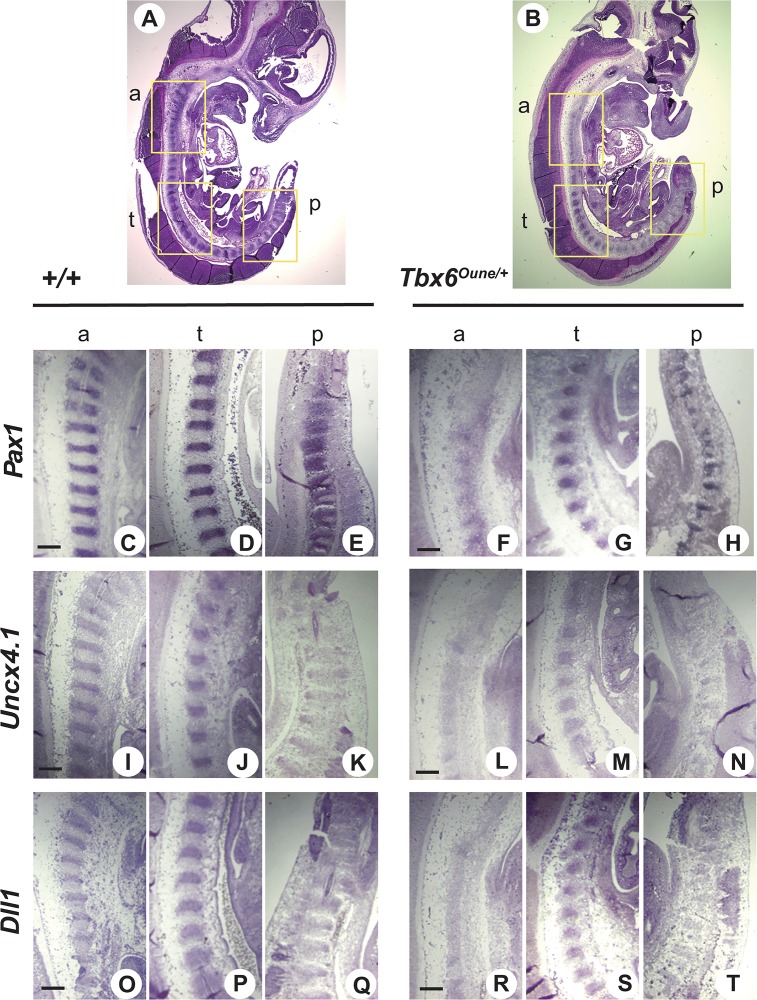
Fig 3. Somite pattering in Oune/+ embryos.
Sagittal sections of E14.5 +/+ and Oune/+ embryos were used for hematoxylin and eosin staining (A, B) and in situ hybridization with various somite markers, Pax1 (C-H), Uncx4.1 (I-N), and Dll1 (O-T). Incomplete somite patterning in the anterior region of Oune/+ embryos was observed (B, box a). In the same embryo, somites in the trunk and posterior regions were morphologically normal (B, boxes t and p). In Oune/+ embryos, Pax1 expression was decreased in the anterior region (F), and somites were dislocated in the posterior region (H). Signals of markers for the caudal half of somites, Uncx4.1 and Dll1, were reduced in the anterior and posterior regions of Oune/+ embryos (L and N: Uncx4.1; R and T: Dll1). Scale bar, 200 μm.

