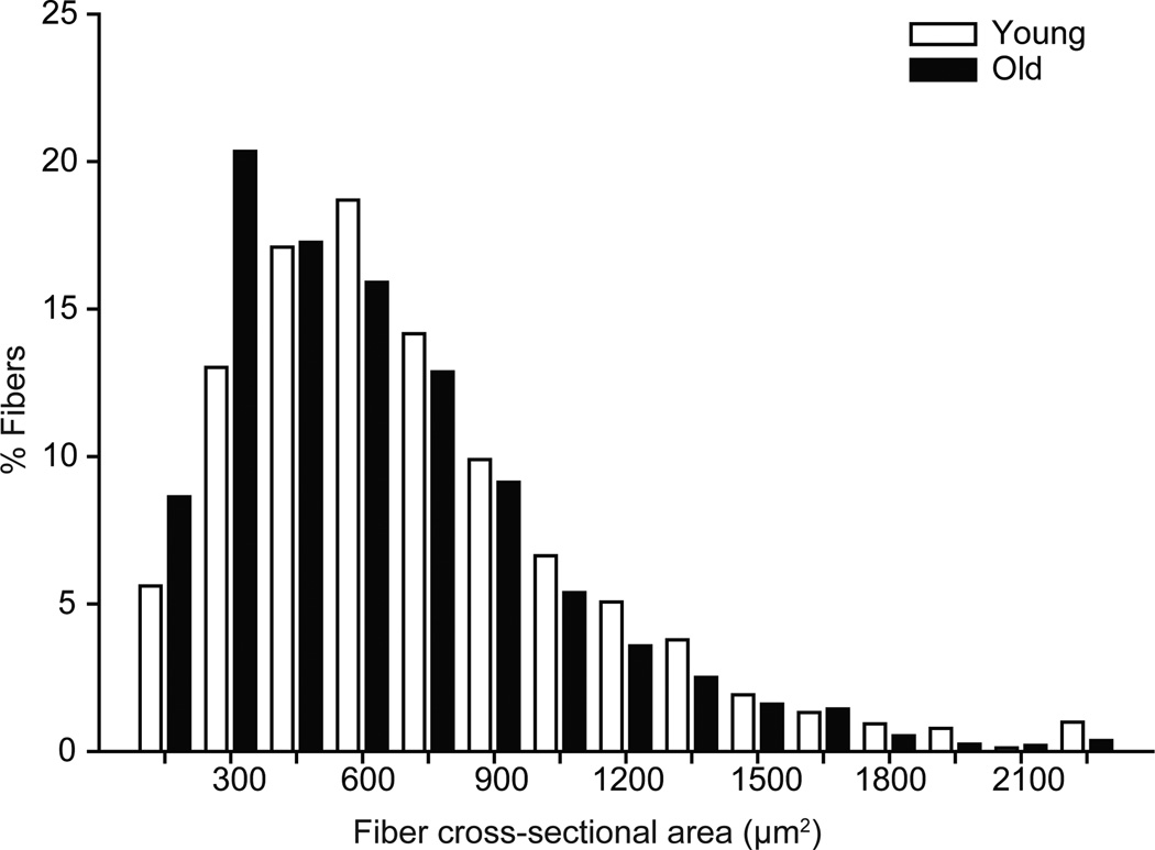
Figure 2.
Diaphragm muscle fiber cross-sectional area from young and old mice, presented as the distribution of fiber cross-sectional areas independent of fiber type. There was a significant left shit in the old fiber cross-sectional areas in comparison to young; distribution was analyzed by chi-squared analysis (P<0.001).

