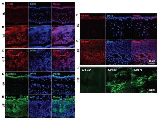
Figure 2. Increased neutrophil infiltration into airway transplants undergoing acute rejection.
(A–C) Neutrophil infiltration, identified by Ly6G staining, into normal tracheas (A), d8 transplants (B) and d12 transplants (C). (D–E) Expression of elastase in normal tracheas (D) and d8 transplants (E). (F–G) Elastase activity in normal tracheas (F) and d8 transplants (G). (H) FITC-lectin perfusion imaging showing that adenovirus-mediated elafin or SPLI transduction protected the microvascular circulation of d12 airway transplants (n=4–6, representative image shown). Scale bars, 20μm (A–G) and 100μm (H).

