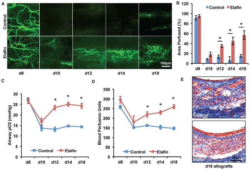
Figure 3. Systemic elafin administration prolongs airway microvascular perfusion, diminishes airway hypoxia and attenuates airway fibrotic remodeling.
(A) FITC-lectin perfusion imaging showing that elafin (1.9mg/kg/d starting on d0) treatment prolongs airway microvascular perfusion. (B) Quantification of perfused areas of the trachea transplant. (C) Airway tissue pO2 measurement following transplantation. (D) Blood perfusion units measured by laser doppler flowmetry. (E) Masson’s Trichrome staining showed that elafin-treated tracheas at d18 display more normal epithelial layer and less subepithelial fibrosis (n=4–6, representative image shown). Scale bars, 100μm (A) and 20μm (E). Data are shown as mean±SEM. Individual time points were compared with the Student’s t test. *P<0.05.

