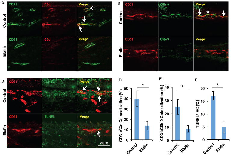
Figure 5. Elafin diminishes complement deposition on airway microvasculature.
(A) Immunofluorescence imaging shows less C3d deposition on ECs (white arrows) in allografts treated with elafin. (B) ECs of allografts treated with elafin have less deposition of C5b-9 complex. (C) EC TUNEL staining shows decreased EC apoptosis in allografts treated with elafin. (D–F) Quantification of C3d deposition (D), C5b-9 deposition (E) and EC TUNEL staining (F) (n=6 per group, representative image shown). Scale bars, 20μm (A–C). Data are shown as mean±SEM. Student’s t test was used to compare groups. *P<0.05.

