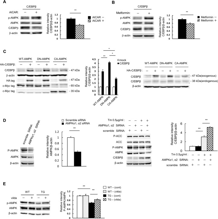
Fig 2. C/EBPβ expression is inhibited when AMPK is activated pharmacologically.
A, B: MIN6 cells were transfected with expression vectors for C/EBPβ. The cells were incubated with 100 μM AICAR (A) or 500 μM metformin (B) for 24 h and analyzed with the indicated antibodies (left). Quantitation of C/EBPβ expression is normalized to β-actin (right). C: MIN6 cells were transfected with expression vectors for c-Myc-tagged AMPK, CA-AMPK, DN-AMPK, and/or human influenza hemagglutinin epitope-tagged C/EBPβ, as indicated. These transfected cells were analyzed with the indicated antibody (left and right). Quantitation of C/EBPβ expression is normalized to β-actin (middle). D: MIN6 cells treated with scramble siRNA (control) and AMPK siRNA were lysed and subjected to immunoblot analysis with antibodies against AMPK, ACC, p-AMPK, p-ACC, and C/EBPβ. E: Isolated islets from WT and TG mice following 8 weeks of vildagliptin (vilda) treatment were analyzed with the indicated antibodies (left). Quantitation of the AMPK phosphorylation level is normalized to total AMPK (right). Data are means ± SE of 3 (C, D) or 4 (A, B, E) independent experiments. *P < 0.05, **P < 0.01.

