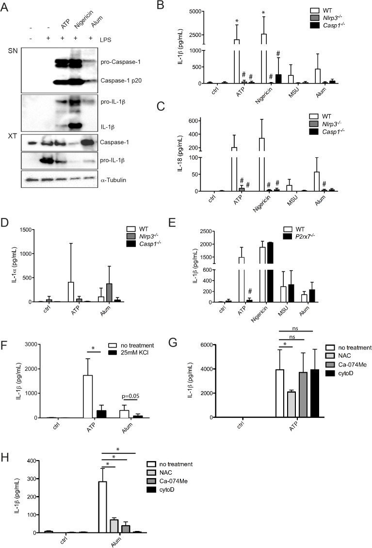
Fig 2. NLRP3 inflammasome activation mechanism in microglia.
LPS primed microglia were stimulated with ATP (1 mM, 30 min), Nigericin (Nig, 1.34 μM, 2 h), Monosodium urate (MSU, 100 μg/ml, 5 h) or Aluminium hydroxide (Alum, 300 μg/ml, 5 h). IL-1β production and caspase-1 cleavage were assessed by Western blot (A). Secretion of IL-1β (B), IL-18 (C) and IL-1α(D) in supernatants of wild-type (WT), Nlrp3 -/- and Casp1 -/- microglia was assessed by ELISA. IL-1β secretion was assessed by ELISA in WT and P2rx7 -/- microglia (E) and after treatment with different inhibitors (F-H) (± KCl (25 mM), N-acetyl cystein (NAC, 5 mM), Ca-074Me (10 μM) and cytochalasinD (cytoD, 2 μM)). Inhibitors were added for 30 min prior to inflammasome activation. Data shown are the mean ± SD of at least three independent experiments. *p<0.05, ns = not significant, compared to control (ctrl) (Fig 3B–3E) or compared to no treatment (Fig 3F–3H), #p<0.05, KO compared to WT.

