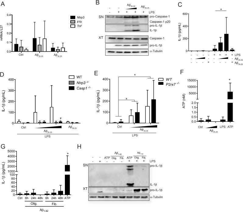
Fig 3. Amyloid-β25–35 activates microglia to secrete IL-1β independently from P2X7 signaling.
Untreated or LPS primed microglia were stimulated with amyloid-β (Aβ)25–35 (20 or 50 μM) or Aβ35–25 (50 μM) for 5 h. (A) RNA was analyzed for expression of Nlrp3, Il1b and Tnf, relative to L27, by Real-Time PCR (B) Cell free culture supernatants (SN) and cell lysates (XT) were analyzed by Western Blot for expression of caspase-1 and IL-1β. α-Tubulin was used as loading control. (C) IL-1β production in culture supernatants was assessed by ELISA. (D, E) IL-1β production in culture supernatants was assessed by ELISA in wild type, Nlrp3 -/- and Casp1 -/- (D), or P2rx7 -/- (E) microglia. (F) ATP release was quantified in cell supernatant upon treatment. (G) Untreated or LPS primed microglia were stimulated with amyloid-β (Aβ) 1–42 (10μM) for 6h, 24h or 48h. IL-1β production in culture supernatants was assessed by ELISA. (H) Cell free culture supernatants (SN) and cell lysates (XT) were analyzed by Western Blot for expression IL-1β. α-Tubulin was used as loading control (I). Data shown are mean ± SD of at least three independent experiments. *p<0.05 compared to control (ctrl), #p<0.05, KO compared to WT.

