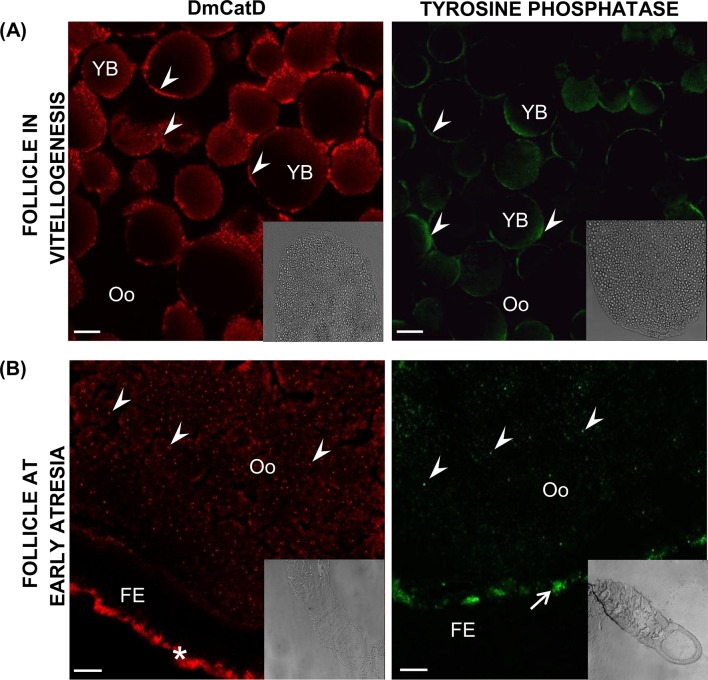
Fig 5. DmCatD and tyrosine phosphatase localization in ovarian follicles by indirect immunofluorescence.
Ovaries from females at vitellogenesis and early atresia were incubated with an anti-cathepsin D antibody (red) or with an anti-tyrosine phosphatase antibody (green) and processed as stated in Materials and Methods. Cryostat sections were analyzed by scanning laser confocal microscopy. (A), in vitellogenic follicles, the fluorescent signal for both acid hydrolases was associated to yolk bodies (arrowheads). (B), at early atresia, oocytes of follicles undergoing incipient degeneration displayed a homogeneous punctate fluorescent pattern for DmCatD and tyrosine phosphatase (arrowheads). At early atresia, DmCatD was also observed in the basal area of follicular epithelium (asterisk), whereas tyrosine phosphatase signal was localized in the perioocytic space of terminal follicles (arrow). Insets correspond to DIC images at lower magnifications. Oo, oocyte; FE, follicular epithelium; YB, yolk bodies. Bars: 10 μm. Similar results were observed after examination of 4–5 ovarioles per ovary in each separate experiment (n = 3).

