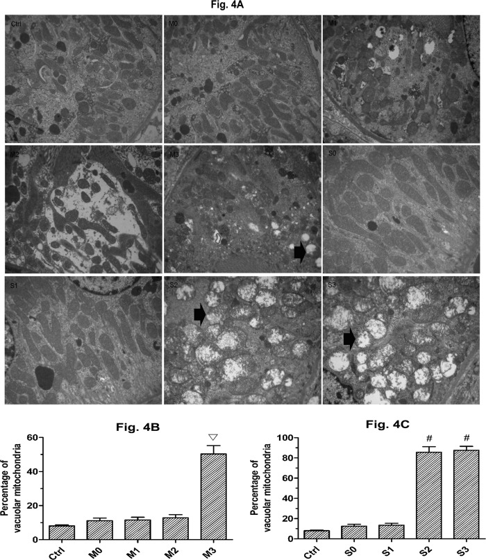
Fig 4. Ultrastructural changes of the mitochondria.
A: Swollen and vacuolar mitochondria in rabbits with mild and severe hydronephrosis under different perfusion pressures (×10000), the arrows showed the swollen and vacuolar mitochondria. B: Percentage of swollen and vacuolar mitochondria in rabbits with mild hydronephrosis. C: Percentage of swollen and vacuolar mitochondria in rabbits with severe hydronephrosis. ▽p<0.05 compared with the M0 group. #p<0.05 compared with the S0 group. Ctrl: normal kidneys perfused with fluid at 20 mmHg, 60 mmHg and 100 mmHg. M0, M1, M2, and M3 represent rabbits with mild hydronephrosis subjected to perfusion pressure of 0 mmHg, 20 mmHg, 60 mmHg and 100 mmHg, respectively. S0, S1, S2, and S3 represent rabbits with severe hydronephrosis subjected to perfusion pressure of 0 mmHg, 20 mmHg, 60 mmHg and 100 mmHg, respectively.

