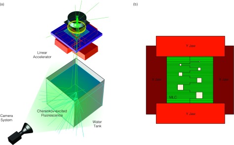
FIG. 1.

In (a), the experimental setup is shown. The camera is placed perpendicular to the radiation beam direction and images the Cherenkov-excited fluorescence through the sidewall of the water tank. In (b), the jaw and MLC configuration that create the field aperture are shown.
