
Fig 2. Revised Fig 1, with additional immunoblots made using a complete replication of the experiment described in the original Fig 1 (including the transfections and lysate preparations) but with samples loaded in the correct consecutive order on the gel.
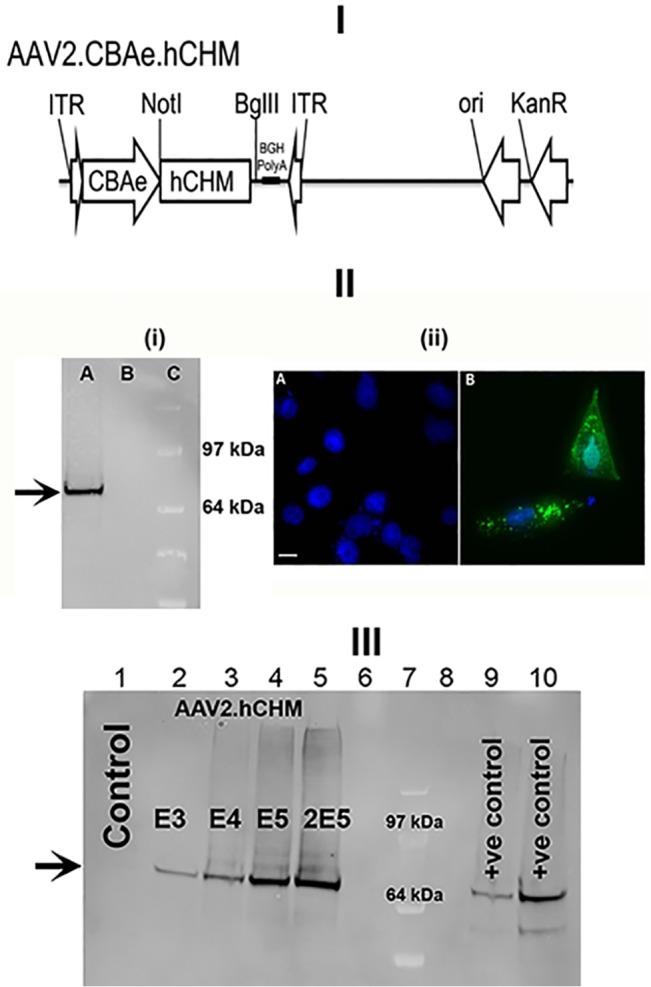
I). Schematic of the AAV proviral plasmid carrying human CHM under the control of the cytomegalovirus enhancer chicken beta actin (eCBA) promoter. ITR: Inverted terminal repeats; Ori: Replication origin; KanR: Kanamycin resistance gene. II) i) Immunoblot and ii) fluorescent analysis reveals REP-1 protein in CHO cells transfected with pAAV2.hCHM. Lane A: Transfected cell (25 μg protein), B: Control (untransfected) cells, C- protein marker (SeeBlue Plus2, Invitrogen, Grand Island, NY). Immunocytochemical analysis revealed the localization of REP-1 to the cytosolic region (II-ii-B; Green). No REP1 is observed in control cells (II-ii-A). Nuclei are stained with DAPI and appear blue. Scale bar is 50 μM. III) Immunoblot analysis of CHO cells infected with 1E3-2E5 viral genomes (vg) of AAV2. hCHM (lanes 2–5) show an increase in REP-1 protein (indicated by arrow) proportional to the titer. Lane 1 is a negative control containing lysate from uninfected CHO cells. Positive (+ve) controls: pAAV2. hCHM-transfected CHO cell lysates (lanes 9, 10). Lanes 6 and 8 were not loaded. Lane 7 contains the SeeBlue Plus 2 protein marker.