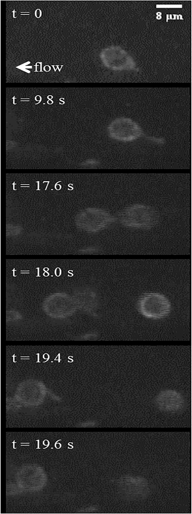
Fig 12. Time-lapse images show a representative, activated neutrophil as it slowly rolls along the vascular wall.

The rolling neutrophil (t = 0 s) is observed to slow down through an extension of a pseudopod attached to the vascular endothelium (t = 9.8 s). It continues rolling by disconnecting the pseudopod to rejoin the flow (t = 17.6 s), reattaches to the endothelium using the same pseudopod (t = 18 s), and finally disconnects the pseudopod (t = 19.4), rapidly retracting it into the cell body and continues to roll along the endothelium (t = 19.6 s).
