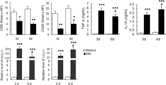
Fig 2. Determination of polarization markers of M.tuberculosis infected MDM.
MDM were infected with M.tuberculosis H37Rv at MOI of 5 and polarization markers were determined at indicated times. A. Phenotypic markers were measured by FCM. MFI was measured and depicted as relative expression as compared to isotype control. Represented is the mean of three donors ±SD. Significance was determined as compared to uninfected group. *P < 0.05, **P < 0.01, ***P < 0.001. B. Effector molecules of macrophages were determined by real-time RT-PCR. Represented is mean ±SD of fold change of mRNA level in infected macrophages to uninfected MDM. C. Cytokines in the supernatant were measured by ELISA. Represented is the mean of three donors ±SD. Significance was determined as compared to uninfected group. *P < 0.05, **P < 0.01, ***P < 0.001.Significance in corresponding marker expression between 6d and 3d was also calculated and represented by the following symbols: #P < 0.05.

