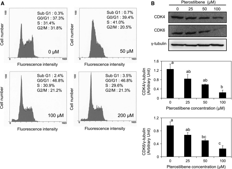
Fig. 3.

The effect of pterostilbene on cell cycle and cell cycle checkpoints protein expression in AH109A cells. a Pterostilbene was dissolved in DMSO and added to the culture medium at final DMSO concentration of 0.1 %. AH109A cells were cultured with medium containing 0, 50, 100, and 200 μM of pterostilbene for 24 h then subjected to cell cycle analysis using flow cytometer. b Effect of pterostilbene on the cell cycle checkpoints protein expression was assessed using western blot analysis. AH109A cells were cultured on medium containing various concentrations of pterostilbene for 24 h then the protein lysates were prepared. Western blot analysis was performed using antibodies to CDK4 (anti-rat CDK4 rabbit monoclonal antibody), CDK6 (anti-rat CDK6 mouse monoclonal antibody), and γ-tubulin (anti-rat γ-tubulin mouse monoclonal antibody). γ-tubulin was used as an internal control. The representative results are shown in the upper panel. The protein bands were quantified by ImageJ and ratios of CDK4/γ-tubulin (middle panel) and CDK6/γ-tubulin (lower panel) are shown. Data are mean ± SEM of 3 independent experiments. Values not sharing a common letter are significantly different by Tukey–Kramer multiple comparison test at p < 0.05
