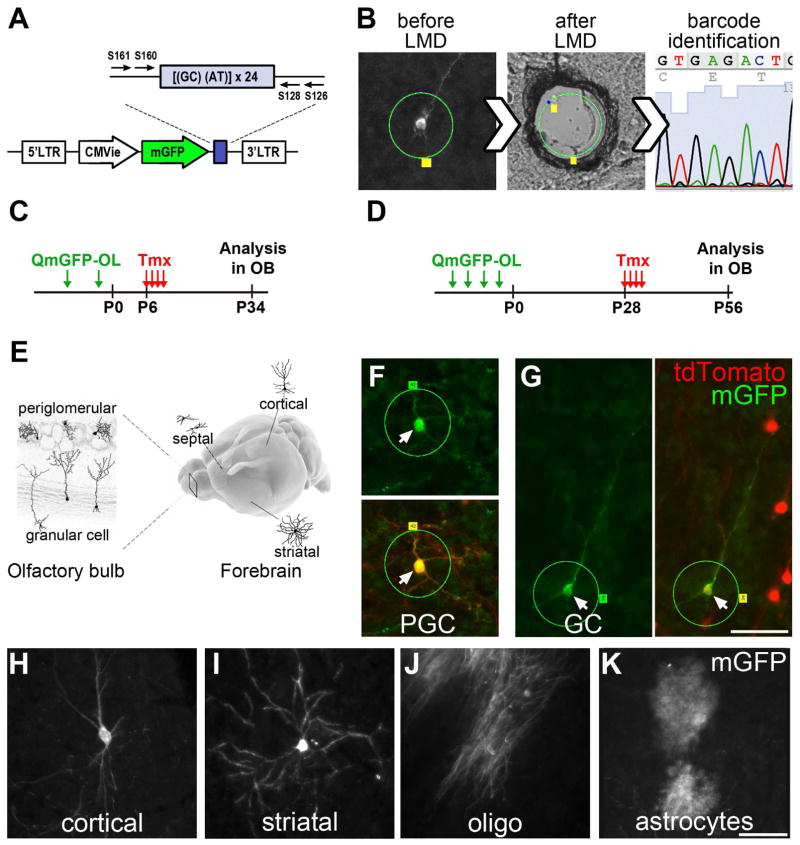
Figure 2. Lineage tracing of individual embryonic progenitors with QmGFP-OL.
A) The QmGFP-OL retroviral library: Each retrovirus expresses mGFP and contains a unique 24 bp sequence. B) M-GFP+ cell tagged for laser microdissection (LMD, green circle), laser-cut and captured, followed by barcode identification. C) QmGFP-OL retroviruses were injected intraventricularly into Nestin::CreER;Ai14 embryos (at E12.5 or E15.5); 6 days after birth, injected mice received Tmx for 4–5d; neurons that were mGFP+/TdT+ in OB and mGFP+ Cx, Hp, St and Sp cells were laser-captured at P34. D) QmGFP-OL retroviruses were injected into Nestin::CreER;Ai14 embryos (at E11.5, E12.5, E13.5 or E15.5); 28 days after birth, injected mice received Tmx; mGFP+/TdT+ OB neurons and mGFP+ Cx, Hp, St and Sp cells were laser-captured at P56. E) Schematics showing regions analyzed and examples of dissected cells in the OB and in Cx, Hp, St and Sp. F–G) Examples of mGFP+/TdT+ PGC (F) and GC (G) before LMD. H–K). Examples of mGFP+ cells found in the telencephalon: neurons (H, cortex; I, striatum), oligodendrocyte (J, cortex) and an astrocyte (K, cortex). Scale bars represent 50 μm in G and K. See also Figure S2A–G.

