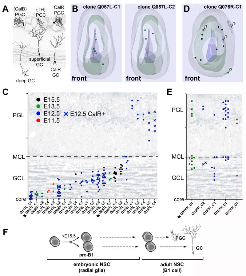
Figure 4. Adult NSCs become specified before E15.5.
A) Main OB interneuron subtypes and their relative layer-specific positions. PGCs are classified by the expression of CalR, TH, and CalB. GCs are classified as deep, intermediate, or superficial, plus CalR expressing. B) Neurolucida® 3D reconstruction of an OB (Q057L) with two P28-generated mGFP+/TdT+ clones: one containing deep GCs (clone Q057L-C1 (circles in left panel) and one containing intermediate GCs (clone Q057L-C2 (circles in right panel). Note that clones are dispersed rostro-caudally, but occupy specific positions in the GC layer (see Movie S1). The OB surface (gray), mitral cell layer (green) and the OB core (blue) are shown. C) The relative OB distances of individual mGFP+/TdT+ cells within clones show that the majority of P28-generated clones contained either one subtype of GC or PGC (39 of 44 multi-cell (≥2) clones; 23 are shown (remaining clones are plotted in Figure 6). D) Neurolucida® 3D reconstruction of one P28-generated clone containing both superficial GCs and PGCs (clone Q076L-C1). Arrows denote PGCs. E) Relative OB distances of individual cells in mixed GC/PGC clones generated at P28 (5 of 44 multi-cell (≥2) clones); these clones contained one GC subtype (superficial or intermediate) and PGCs. Clones containing deep and superficial GCs were not encountered in any of our embryonic injections. F) Pre-B1 cells are specified for the production of GCs or PGCs subtypes during early embryonic development before E15.5 (see text). Closed circles and crosses denote CalR negative and positive cells, respectively. Asterisks mark Neurolucida® reconstructed clones in C and E. GCL: granule cell layer, MCL: mitral cell layer, PGL: periglomerular cell layer. See also Figure S2H–J and Table S3.

