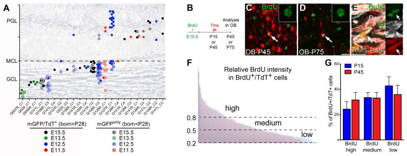
Figure 6. Pre-B1 cells remain quiescent until their reactivation in the postnatal brain.

A) Relative OB positions of mGFP+/TdT+ (filled circles, born after P28) and mGFPonly (open circles, born before P28) cells within clones (experiment in Figure 3 D–E). Eleven out of 23 mGFP+/TdT+ OB clones were clonally related to mGFPonly OB neurons and of these 4 were mixed suggesting an origin in the embryo (see also Table S4). B) E15.5 pregnant Nestin::CreER;Ai14 females were BrdU injected; littermates received Tmx at either P15 or P45 and their OBs were analyzed 28d later. C–E) Independent of the time of Tmx administration, brightly labeled BrdU+ nuclei were detected in TdT+ OB neurons (C, D) and TdT+/DCX+ neuroblasts in the RMS (E), indicating limited BrdU dilution. F) Distribution of the relative BrdU intensities in BrdU+/TdT+ OB neurons from both age groups (blue: Tmx P15, n=132 cells; red: Tmx P45, n=126 cells. 7 mice/group, three independent experiments). (G) Percentages of TdT+ OB neurons with high, medium or low BrdU intensities in P15 (blue) or P45 (red) Tmx-treated mice. Data are expressed as mean ± s.e.m., n=7 mice/group, three independent experiments each (n.s.). GCL: granule cell layer, MCL: mitral cell layer, PGL: periglomerular cell layer. See also Table S4.
