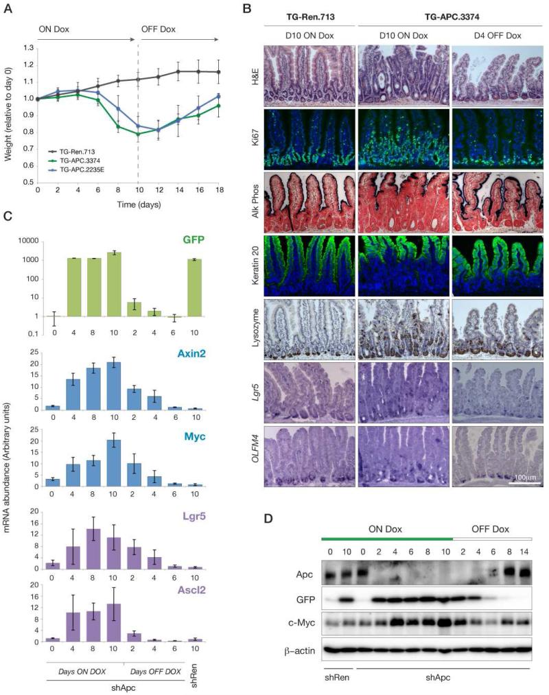
Figure 1. Acute depletion of Apc in the mouse.
A. Animal weight during dox treatment and following dox withdrawal at 10 days, normalized to Day 0 within each cohort. Lines represent TG-Ren.713 (black), TG-Apc.3374 (green) and TG-Apc.2235E (blue) mice. B. Immunohistochemical (H&E, Alkaline phosphatase and Lysozyme), immunofluorescent (Ki67 and Keratin 20) stains and in situ hybridizations (Lgr5 and Olfm4) from shRen (control) and shApc intestine following dox treatment for 10 days (left two panels) and withdrawn from dox for 4 days (right panel). C. Quantitative RT-PCR analysis of gene expression in intestinal villi following dox treatment and withdrawal for TG-Apc.3374 mice as indicated. Markers of transgene induction (GFP), Wnt activation (Axin2, Myc) and stem cells (Lgr5, Ascl2) are shown for each time point. Gene expression in day 10-treated TG-Ren.713 is indicated to the right on each plot. D. Western blot of whole cell lysates from intestinal villi following dox treatment and withdrawal at time points indicated, for TG-Ren.713 and TG-APC.3374 mice, probed for Apc, GFP, cMyc and β-Actin, as indicated.

