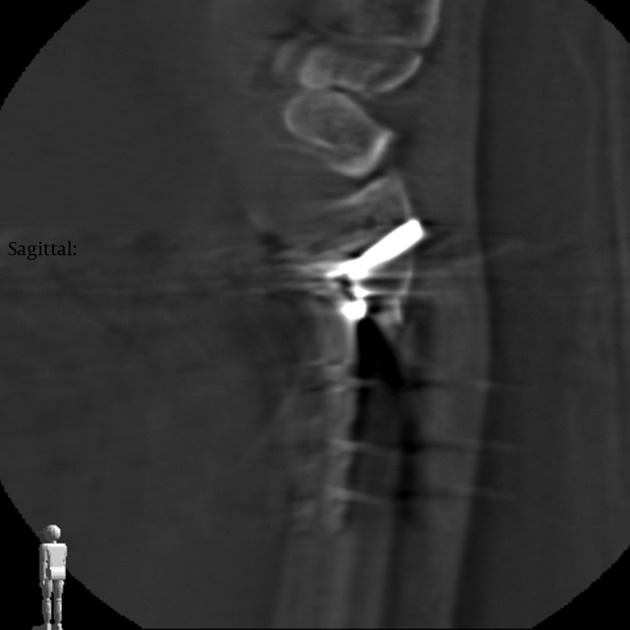
Figure 2. Two-Dimensional Reformation Images of the Intraoperative Three-Dimensional Fluoroscopic Scan After Volar Distal Radius Plating.

In the sagittal plane, perfect juxta-articular screw placements are displayed. In the coronal plane, a screw tip perforation was detected (the image quality in the axial plane is limited).
