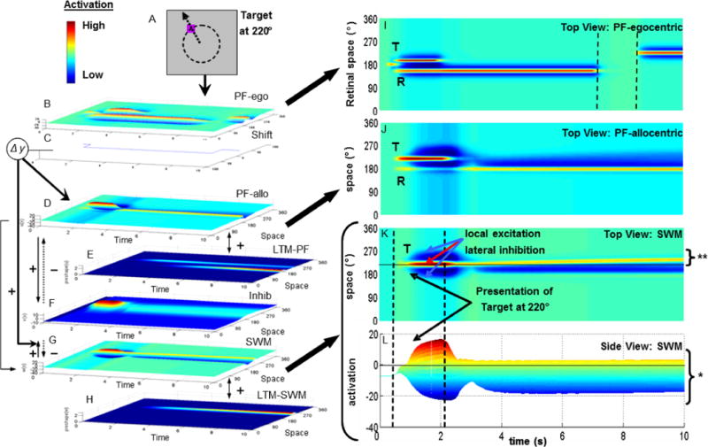
Figure 1.

DFT simulation of one spatial recall trial with target at 220° (see dashed circle and arrow in A; 180° = midline of task space). Model architecture (B–H) consists of seven layers of spatially-tuned neurons with time shown on the x-axis, activation on the y-axis (see color inset), and space on the z-axis. Arrows between layers indicate excitatory (solid) and inhibitory (dotted) projections.
Panels in the right column show top view of three fields. The egocentric perceptual field (PF-ego; B) is shown in I with time along the x-axis and space along the y-axis. Input is presented to this field in retinal coordinates. Two activation peaks (red activation) are present at the start of the trial corresponding to the target (T), which disappears, and the reference axis (R), which remains visible. Dashed lines indicate a brief occlusion of the reference (i.e., no input). After the occlusion, the reference shifts from 160° to 230° in the retinal frame.
The allocentric perceptual field (PF-allo; D) is shown in J (axes as in I). Note that the reference peak (R) remains active and centered at 180°, even though it was occluded and shifted in PF-ego. This is because the shift field (C) transforms input from the egocentric to the allocentric frame. The spatial working memory field (SWM; G) is shown in K (axes as in I) and L (activation along the y-axis). The target peak (T) sustains after input is removed (* in L), and drifts away from midline over delay (** in K; see ‘bending’ yellow line).
