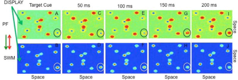
Figure 4.

Fine-grained view of the loss of a SWM peak during tracking. 4A shows the cuing of 6 targets which are maintained in SWM (see blue inhibitory troughs in PF and red activation peaks in SWM). Across 4C–F, SWM updates the location of all 6 targets. However, after 100 ms of tracking the circled item in 4F has weakened. By 150 ms, the circled SWM peak spontaneously decays (4H). Consequently, the previously tracked item becomes a distractor, which, in turn, leads to an error at the end of the trial.
