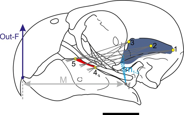
Figure 1.

Biomechanical modelling of the jaw in Myiopsitta monachus. Two lines between the most anterior (5) and posterior (4) part of the insertion and the most posterior (1), center (2) and most anterior part (3) of the origin are drawn. The angle between the two lines is subdivided into several lines of action. All the moment arms (m1-3) are measured for each point (posterior, center and anterior) and a mean moment arm is estimated for each point. With the three moments arms, a mean moment arm is calculated. The procedure is then repeated for other muscles (input-force). See text for further explanation. Abbreviations: 1–3, most posterior, center and anterior points of the muscle origin; 4–5, most posterior and anterior edges of muscle insertion; f, fulcrum; m1-3, in-lever moment arms for the three lines between points 1 and 4 and 5; M, out-lever moment arm; out-F, out-force. Scale = 1 cm.
