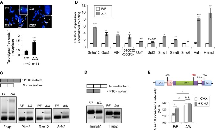
Figure 3. Smg6 is required for telomere maintenance and NMD in ESCs.
A Telomere FISH analysis of control (F/F) and Smg6Δ/Δ (Δ/Δ) ESC metaphases. Representative images (up) and quantification of chromosomes lacking telomere signals (telomere-signal-free ends) (lower panel) are shown. n, the number of metaphases analyzed.
B qRT–PCR analysis of NMD target transcripts in control and Smg6Δ/Δ ESCs. The expression levels of the NMD target genes were normalized to β-actin. The data are from three independent biological samples.
C RT–PCR analysis of exon inclusion generated PTC-containing (*PTC+) isoforms in control and Smg6Δ/Δ ESCs.
D RT–PCR analysis of exon exclusion generated PTC-containing (*PTC+) isoforms in control and Smg6Δ/Δ ESCs.
E Smg6Δ/Δ ESCs are NMD defective. ESCs were transfected with the NMD reporter (upper panel) and analyzed in triplicate by FACS. The GFP signal intensity determines the NMD activity. CHX was used to inhibit NMD. The data represent one of two independent ES clones of each genotype.
Data information: The error bars represent the SEM. Unpaired Student's t-test was used. n.s., not significant, P > 0.05; *P < 0.05; **P < 0.01; ***P < 0.001.Source data are available online for this figure.

