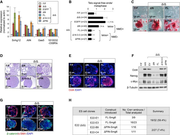
Figure 4. Smg6-mediated NMD function determines ESC differentiation.
A qRT–PCR assay of NMD target gene expression after reconstitution of Smg6Δ/Δ ESCs with NMD-proficient or NMD-deficient Smg6 constructs (see Supplementary Fig S5A). The data are from three independent qRT–PCR experiments.
B Telomere FISH analysis of metaphases of ESCs with indicated genotype and reconstituted with indicated Smg6-expression vectors. The number of telomere-signal-free ends per metaphase is shown. n, the total number of metaphases used for the quantification. NMD+, NMD-proficient vectors; NMD-, NMD-deficient vectors; telos+, telomere-proficient vectors; telos-, telomere-deficient vectors. Statistic analysis was used to compare the Δ/Δ samples and their derived clones with different Smg6 truncation expression vectors.
C Spontaneous differentiation of Smg6Δ/Δ (Δ/Δ) ESCs reconstituted with NMD-proficient (FL and ΔN) or NMD-deficient (Δ14-3-3 and ΔPIN) Smg6 constructs. Representative images of AP staining of cultures at day 6 after the removal of LIF and feeder are shown.
D EB formation assay of Smg6Δ/Δ ESCs reconstituted with NMD-proficient or NMD-deficient Smg6 vectors. The morphology of EB cells and cavity formation at day 8 are indicative of the differentiation status.
E Immunofluorescence analysis of expression of the stem cell marker Oct4 on EBs of reconstituted Smg6Δ/Δ ESCs on day 8.
F Western blotting of Smg6Δ/Δ EB cells reconstituted with various Smg6 truncation vectors on day 8. The expressions of Oct4, Nanog, and c-Myc are shown. β-Tubulin was used as a loading control.
G Immunofluorescence analysis of expression of markers β-catenin and α-SMA on EBs of reconstituted Smg6Δ/Δ ESCs on day 8.
H Chimerism analysis of in vivo rescue of differentiation defects of Smg6Δ/Δ ESCs after reconstitution with NMD-proficient (FL-Smg6) or NMD-deficient (ΔPIN-Smg6) construct. Parental ES clone E22 (Smg6-CER) contained the Cre sequence, which was used to detect ESC contribution in E12.5 embryos by PCR. All ESC clones were pretreated with 4-OHT for 6 days to delete endogenous Smg6 before blastocyst injection.
Data information: The error bars represent the SEM. Unpaired Student's t-test was used. n.s., not significant; **P < 0.01. Source data are available online for this figure.

