
Figure 6.
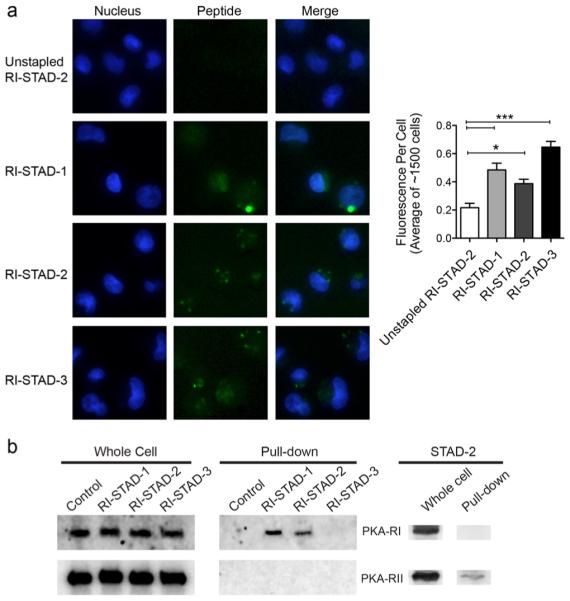
RI-STAD peptides are cell-permeable and selectively bind the RI isoform in cells. (a) Fluorescent images and quantification of live cells after treatment with FITC-labeled peptides (5 μM) for 6 h shows that RI-STAD-1, -2, and -3 demonstrate enhanced intracellular localization in cells. Quantification was performed using 28–32 fields (n = 1410–1670 cells). * p < 0.05, *** p < 0.001 relative to the unstapled control. (b) RI-STAD-1 and RI-STAD-2 bind the RI isoform, but not RII, in cells. MDA-MB-231 cell were treated with 5 μM biotin-labeled peptides for 12 h before lysis. Pulldowns were performed using avidin-coated resin, and the RI and RII isoforms were detected by immunoblotting. As a control, the peptide STAD-2 binds only the RII isoform, not RI.