
Figure 3. Distinct Mechanisms of RIG-I and MDA5 Filament Formation.
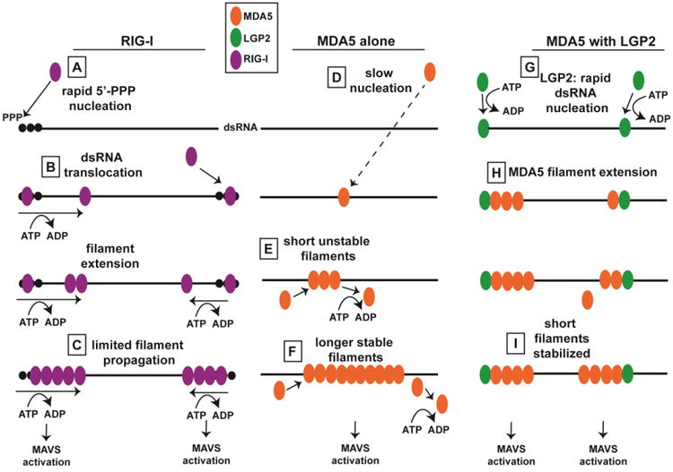
Figure illustrates distinct mechanisms of RIG-I and MDA5 filament formation, and depicts how LGP2 may regulate MDA5 filament assembly. (A) RIG-I CTD recognizes 5′-triphosphorylated dsRNA. (B) ATP-dependent dsRNA translocation by RIG-I aids in filament assembly, leading to limited filament propagation (C). MDA5 nucleation occurs very slowly (D), and short filaments are unstable (E), while filament extension leads to stabilization via protein-protein interactions on adjacent MDA5 monomers (F). LGP2 may nucleate MDA5 filaments by binding dsRNA termini (G). MDA5 filaments extend (H), and LGP2 stabilizes the formation of these shorter filaments (I).