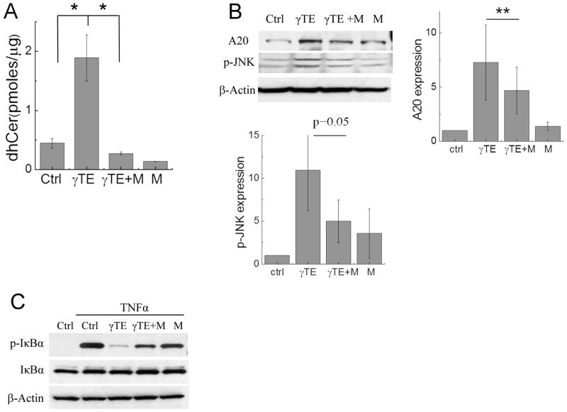
Figure 6. Myriocin reversed γTE-caused increase of dhCer, induction of A20 and subsequent inhibition of NF-κB.
Panels A and B: RAW264.7 cells were incubated with γTE (20 μM) for 8 h (A) or 14 h (B) in the presence or absence of 6 μM myriocin (M). Total amounts of dhCers were measured by LC-MS/MS (A). Cytosolic proteins (for p-JNK) or whole proteins (for A20) were analyzed by Western blot that was quantified by ImageJ (B). Panel C: RAW cells were incubated with γTE (20 μM) for 14 h with or w/o 6 μM myriocin (M) and then were stimulated with TNFα for 5 min. Cytosolic proteins were used for monitoring IκBα phosphorylation. **P < 0.01 indicates significant difference between γTE and γTE plus myriocin (γTE+M) treated cells (Mean ± SEM, n = 3–4).

